|
---------------------------- 2025 Platform Presentation Winner --------------------------
DNA Methylation Profiling of Uterine
Mesenchymal Tumors Suggests Adenosarcomas
Are a Heterogeneous Group of Neoplasms
Minami A. Tokuyama, Erna Forgo, Emanuela D'Angelo, Reem Al-Humadi, Leandra G. Kingsley, Arslan Kasimov, Sabrina Zdravkovic, Sarah Lawson, Tamra-Lee McCleary, Jennifer A. Bennett, Carlos Parra-Herran, David L. Kolin, Marisa Rose Nucci, Charles Matthew Quick, Glenn McCluggage, Brooke E. Howitt
Background: Uterine mesenchymal tumors (UMT) are rare neoplasms
with overlapping morphologic features and
immunophenotypes. In some cases, molecular features
are entity-defining (such as YWHAE or ALK fusion), and
testing may be necessary for accurate diagnosis. Uterine
tumors resembling ovarian sex cord tumors (UTROSCT)
display varying degrees of sex cord differentiation (SCD)
and harbor frequent NCOA-fusions, differentiating them
from most other tumors with SCD. Mullerian
adenosarcomas (AS) are malignant UMTs with associated
non-neoplastic epithelial components and can also have
extensive SCD. No unifying molecular features have been
identified in AS, but a subset of these tumors (11%) also
harbor NCOA-fusions.
Design: We collected a multicenter cohort of 169 UMTs other than
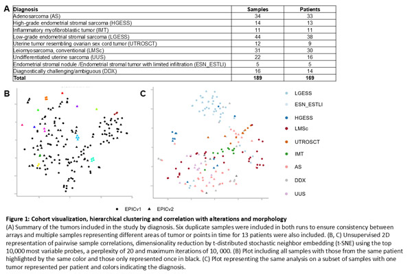
benign leiomyomas, including 34 AS (Fig 1A). DNA was
extracted from formalin-fixed paraffin embedded tissue.
Methylation data was generated using the Infinium
MethylationEPIC v2.0 or v1.0 BeadChip arrays.
Unsupervised hierarchical clustering was performed using
the top 10,000 probes with the highest median absolute
deviation across beta values using Euclidean distance
squared and Ward's linkage method. Morphologic
features and any known molecular alterations were
recorded when available.
Results: Samples run on both the v1.0 and v2.0 arrays (n=6) as
well as tumor samples with different locations/timepoints
from the same patient (n = 13) showed high concordance
(Fig 1B). LGESS and HGESS clustered separately (Fig
1C). Other tumors in diagnostic categories with known
recurrent molecular alterations such as UTROSCT and
IMT tended to cluster by diagnosis. In contrast, tumors
from diagnostic categories without unifying molecular
alterations such as AS were not cluster-exclusive. Most
AS clustered into 1 of 3 groups: 1) 5 AS clustered with
UTROSCT; 2 exhibited SCD, none had sarcomatous
overgrowth (SO), and all were low-grade, 2) 7 AS with SO
clustered with UUS and 3) 12 AS formed a group with
JAZF1-rearranged LGESS and UMTs with ambiguous
morphologic features that lacked sequencing data (Fig 2).

Conclusions: Our results suggest some UMT groups, especially AS,
have heterogenous underlying biology. Specifically, we
confirm the reported molecular similarities between
UTROSCT and a subset of AS. Though our study is
limited, the data warrant further investigation of
molecular-based classification schemes and demonstrate
added benefit of methylation profiling in UMTs.
|